
 Call Us:
011 4338 8888
Call Us:
011 4338 8888

 Book an Appointment
 Locate us on Google Map
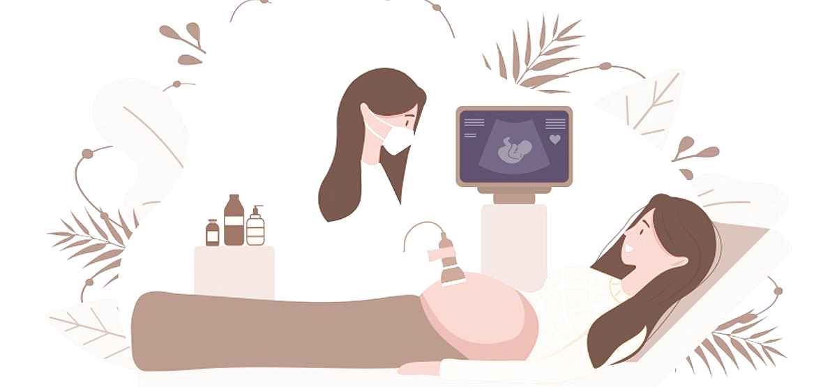
A foetal ultrasound (also called a sonogram) uses sound waves to produce images on a monitor of a foetus inside the uterus. Foetal ultrasounds can greatly help your health care providers examine and assess your baby's growth, and your health and monitor your pregnancy better. Also, in other cases, foetal ultrasounds are used for evaluating any potential problems or confirming a diagnosis.
If you’re pregnant or planning one, you should know that ultrasounds are safe and if you wish to get the best ultrasound in Dwarka, you can visit us today and consult with some of Delhi’s best gynaecologists at Aakash Healthcare.
The first foetal ultrasound is done in the first trimester to confirm your pregnancy and also to get an estimate on how much time has passed since you’ve been pregnant. If your pregnancy is uncomplicated, the next ultrasound is slated to be during the second trimester, when anatomic details become visible. If somehow a problem is discovered, you may need to have another ultrasound or get additional imaging tests, such as MRIs, which may be recommended.
There are two main types of foetal ultrasound tests:
As it turns out, most women require just a handful of ultrasounds during pregnancy. Read on to learn more about these important pregnancy scans you’ll take and for which trimester.
In a pregnancy ultrasound, a doctor or highly skilled technician will use a plastic transducer to transmit high-frequency sound waves through your uterus, after which these sound waves send signals back to a machine that converts and displays them in the form of images of your baby.
Ultrasounds do provide us doctors with a raft of valuable information—for instance, they help monitor the baby's growth, detect abnormalities, determine if you have twins or more, predict your due date, show the position of the placenta, and can also indicate the sex of your baby (this is prohibited in India).
To prepare you for all the important pregnancy scans you’ll need, we’ve broken down the most common types of ultrasounds there are and when you’ll have to get them.
The first ultrasound, also called a baby sonogram, might take place when you're about six to eight weeks pregnant. However, every woman will not need to get this scan; doctors mostly conduct it for high-risk pregnancy complications like bleeding, abdominal pain, and particularly if there is a history of miscarriage or birth defects.
An early pregnancy ultrasound can be done transvaginally so doctors get a clear picture of the baby
The process has been discussed above.
At about six weeks gestation, we can begin to see the baby's heartbeat and your gynaecologist will also be able to predict your baby's due date, keep track of milestones, determine the number of babies in the womb, and detect an ectopic pregnancy.
Those who don’t get the first six to eight-week ultrasound can have a"dating ultrasound" after about weeks 10 to 13 of pregnancy. This will give parents the same information: due date, the baby's "crown-rump length" (measurement from head to bottom), the number of babies inside the womb, and the baby’s heartbeat.
Between 14 and 20 weeks, you could also get a nuchal translucency test (NTT) to check for Down syndrome, any heart defects, or other chromosomal abnormalities. Women should also consider getting this if their initial screening tests have revealed any potential problems, if they're 35 or above, or if they have a family history of certain birth defects.
In a nuchal translucency screening, a doctor uses an ultrasound to assess the thickness at the back of the baby's neck (they also measure hormones and proteins through a blood test). A thicker than normal neck can indicate an increased risk of birth defects and trisomy 18.
This is a detailed pregnancy ultrasound, which happens in the middle of weeks 18 and 20 in the second trimester, and lasts about 20 to 45 minutes if you're carrying one baby and longer if you're carrying multiples. This scan is the most thorough check-up you and your baby will have before they're born.
In an anatomy scan, a doctor will check the baby's heart rate and look out for any abnormalities in the brain, kidneys, heart, and liver. Doctors also count the baby's fingers and toes, examine the placenta, look for birth defects, and measure the amniotic fluid level.
The baby’s sex is also more or less determinable by now.
Most parents in waiting don't need an ultrasound in the third trimester unless their pregnancy is high-risk. In case you have high blood pressure, bleeding, or low levels of amniotic fluid, or if you’re over 35 —a doctor may choose to perform in-office, low-resolution ultrasounds during your visits for reassurance. You'll also need to get a follow-up ultrasound if your cervix was covered by the placenta at the time of the 20-week scan.
Also, Read: Understanding an Ultrasound and its purpose, the procedure, and use
 011 4338 8888


Leave a Reply
Your email address will not be published. Required fields are marked *